KNOCK KNEES/GENU VALGUM
Knock knees (genu valgum) is a condition where the knees tilt inward and touch each other while the ankles remain apart. In majority of cases, this is a normal part of a child's growth and development.
1. The Natural Timeline of Leg Growth
Orthopaedic surgeons often describe the way a child's legs grow as a "pendulum" that swings from bowed to knock-kneed before settling into a straight alignment.
Birth to 18–24 months: Most babies are bow-legged (genu varum). This is normal "packaging" from the womb.
Ages 2 to 5: The legs naturally shift into a knock-kneed position. This usually peaks around age 3 or 4.
Ages 7 to 8: The legs typically straighten out on their own. Most children (99%) will have normal alignment by this age.
2. Is Treatment Necessary?
For the vast majority of children, no treatment is required.
Observation: The standard "treatment" is simply watching the child grow. Doctors may measure the distance between the ankle bones (intermalleolar distance) to track improvement over time. Monthly walking videos and standing photographs can also be clicked for monitoring.
Outdated Methods: Special shoes, wedges, shoe inserts, or daytime braces do not work and are no longer recommended. They do not speed up the natural straightening process and rather add to the morbidity of the child.
Activity: Children with knock knees do not need to limit their physical activity; they can run, jump, and play sports like any other child.
3. When to See a Specialist (Red Flags)
While most cases are "physiologic" (normal), some are "pathologic" (caused by an underlying issue). Parents should consult a paediatric orthopaedic surgeon if they notice any of the following:
| Red Flag | Why it Matters |
|---|---|
| One-Sided (Unilateral) | If only one knee turns in, it may indicate a previous injury to the growth plate or a bone infection. |
| Severe Gap | Ankle separation of more than 8 cm (approx. 3 inches) when the knees are touching. |
| Pain or Limping | Normal knock knees should not be painful. Pain or a limp suggests a structural problem. |
| Short Stature | If the child is also very short for their age, it could signal a metabolic issue such as rickets or a skeletal dysplasia. |
| Age | Knock knees that appear for the first time before age 2 or after age 7, or that worsen after age 8. |
4. Surgical Options (Rare Cases)
If the condition is severe, painful, or persists into late childhood/adolescence, a surgeon may recommend:
Guided Growth (Hemiepiphysiodesis): A minimally invasive procedure where a small metal plate or screw is placed on the inner side of the growth plate. This "tethers" the growth on one side, allowing the other side to grow and naturally straighten the leg. This must be done while the child is still growing.
Osteotomy: For older teens whose growth plates have closed, the bone may need to be cut and realigned. This is a much more significant surgery and is reserved for the most severe cases.
Summary for Parents
In most cases, knock knees are a "cosmetic" phase of childhood that will disappear by the time your child reaches second or third grade. If your child is growing well, has symmetric legs, and is not in pain, the best medicine is usually just time.
BOWLEGS
Bowlegs (genu varum) is the physical opposite of knock knees. It is characterized by the knees staying wide apart while the feet and ankles are touching.
Just like knock knees, most cases are a normal part of development, but because it is the first stage in a child's leg growth, it is often the first thing parents notice.
1. The Growth Cycle (The "S-Curve")
The legs go through a predictable transformation as a child matures:
0–18 Months (Bowlegs): Almost all babies are born bowlegged due to their cramped position in the womb. This is called Physiologic Genu Varum.
18–24 Months (Straightening): The legs naturally begin to straighten as the child starts walking and bearing weight.
2–5 Years (Knock Knees): The "pendulum" swings the other way, and most children become knock-kneed.
Age 7+ (Adult Alignment): The legs settle into their permanent, slightly straight-to-knock-kneed adult position.
2. Is it Normal or a Problem?
POSNA distinguishes between Physiologic (Normal) and Pathologic (Medical) bowlegs.
| Feature | Physiologic (Normal) | Pathologic (Worrying) |
|---|---|---|
| Symmetry | Both legs are bowed equally. | One leg is bowed significantly more than the other. |
| Age | Most common under age 2. | Persists or gets worse after age 2. |
| Walking | Child walks and runs normally. | Child has a noticeable limp or “lateral thrust” (knee jerks outward). |
| Height | Child is growing at a normal rate. | Child is unusually short for their age. |
3. Medical causes to Rule out
If the bowing doesn't resolve by age 2, a specialist will look for these conditions:
Blount’s Disease: A growth disorder of the shinbone (tibia) that causes the bowing to worsen rather than improve. It is more common in children who are overweight or who walked very early (before 11 months).
Rickets: A softening of the bones usually caused by a severe deficiency in Vitamin D or Calcium.
Skeletal Dysplasia: Genetic conditions that affect how bones and cartilage grow.
4. Treatment Facts
What NOT to do: POSNA and OrthoKids explicitly state that special shoes, wedges, and nighttime splints do not work for physiologic bowlegs and are not recommended.
Observation: If the child is under age 2 and symmetric, the doctor will likely just measure the distance between the knees and see you back in 6 months.
Bracing/Surgery: These are only used for pathologic conditions like Blount’s Disease. Surgery (Guided Growth) is usually a last resort if bracing fails or if the child is too old for a brace.
Summary Table: Bowlegs vs. Knock Knees
| Bowlegs | Knock Knees | |
|---|---|---|
| Appearance | Knees apart, ankles together. | Knees together, ankles apart. |
| Peak Age | 0 to 18 months. | 3 to 5 years. |
| Self-Correction | Usually by age 2. | Usually by age 7 or 8. |
| Measurement | Space between the knees. | Space between the ankles. |
Cases - Videos
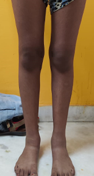
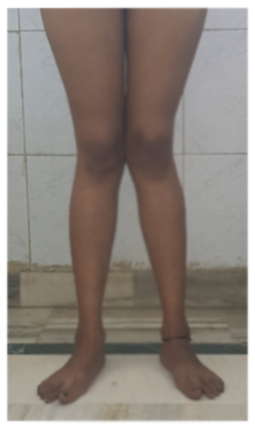
Case - 1 (Knock Knees)
Before
Pre-operative clinical image showing bilateral genu valgum (Left more than right).
After
6 months post operative image showing well corrected left lowerlimb. Patient is yet to be operated for the right side.
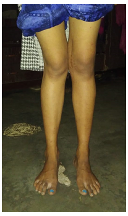
Case - 2 (Knock Knees)
Before
After